12.1 Introduction
If somebody thinks they may have broken a bone, a doctor may request an X-ray image be taken. An X-ray image, or its 3-D counterpart the CAT scan, is useful for looking at bones. The calcium in the bones absorbs the X-rays well and does not let them pass through the bones and reach the detector. But, if a doctor wants to get an image of organic tissue in the patient, X-rays do not work very well. Instead, the doctor may request an MRI (magnetic resonance imaging) for the patient. In an MRI, the patient is inserted into a tube that is surrounded by a very powerful magnet. At the same time, invisible, silent, and harmless radio waves are sent towards the patient. This combination of a powerful magnet and radio waves allows the doctor to look at the concentration of nuclei of hydrogen atoms in the organic tissue. Thankfully, our organic tissue is full of hydrogen atoms.
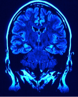
Photo by jkt_de at Morguefile.com
Using a technique that is nearly identical to an MRI, chemists can insert a chemical compound into a small tube surrounded by a powerful magnet. Radio waves are directed at the sample and information about the location of the hydrogen atoms on the molecule is obtained. Chemists call this technique nuclear magnetic resonance, NMR. Unfortunately for chemists, our pictures are not as nice to look at as what doctors get with an MRI. A chemist’s NMR spectrum has information, but it ends up being a little puzzle that needs solved to determine how many and where the hydrogen atoms are on a molecule. It is called “nuclear” magnetic resonance because the magnet affects the atomic nuclei and not the electrons of the atoms. In common everyday language, the word “nuclear” invokes thoughts of nuclear bombs, radioactivity, and danger. At one time, doctors would use nuclear magnetic resonance (NMR) on patients. But, since patients were unnecessarily avoiding the procedure because they were frightened about radioactivity, the medical establishment changed the name of this diagnostic tool to MRI to avoid the “nuclear” fear. By the way, even though X-rays and CAT scans do use high-energy light and do slightly increase the chances of causing mutations in cells and thus cancer, there is no chance that an MRI can do so. Radio waves are very low energy and cannot damage DNA.